発生原理に基づくヒト神経筋接合部モデルの開発 —発生領域を操作できるヒト神経・筋創薬プラットフォームの構築—

元野 誠
MOTOMO MAKOYO

医学部 解剖学教室


カテゴリー:医薬品・医療素材
 

研究開発段階


概要

脊髄運動ニューロンと骨格筋の共通の前駆細胞である神経中胚葉前駆細胞(NMPs) を起点に、同一培養系で運動ニューロンと骨格筋を同時に誘導することで、 ヒト人工多能性幹細胞(iPS細胞)から、マウスでは再現できないヒトの発生原理に基づいた神経筋接合部(NMJ)モデルを構築した。

技術の特徴

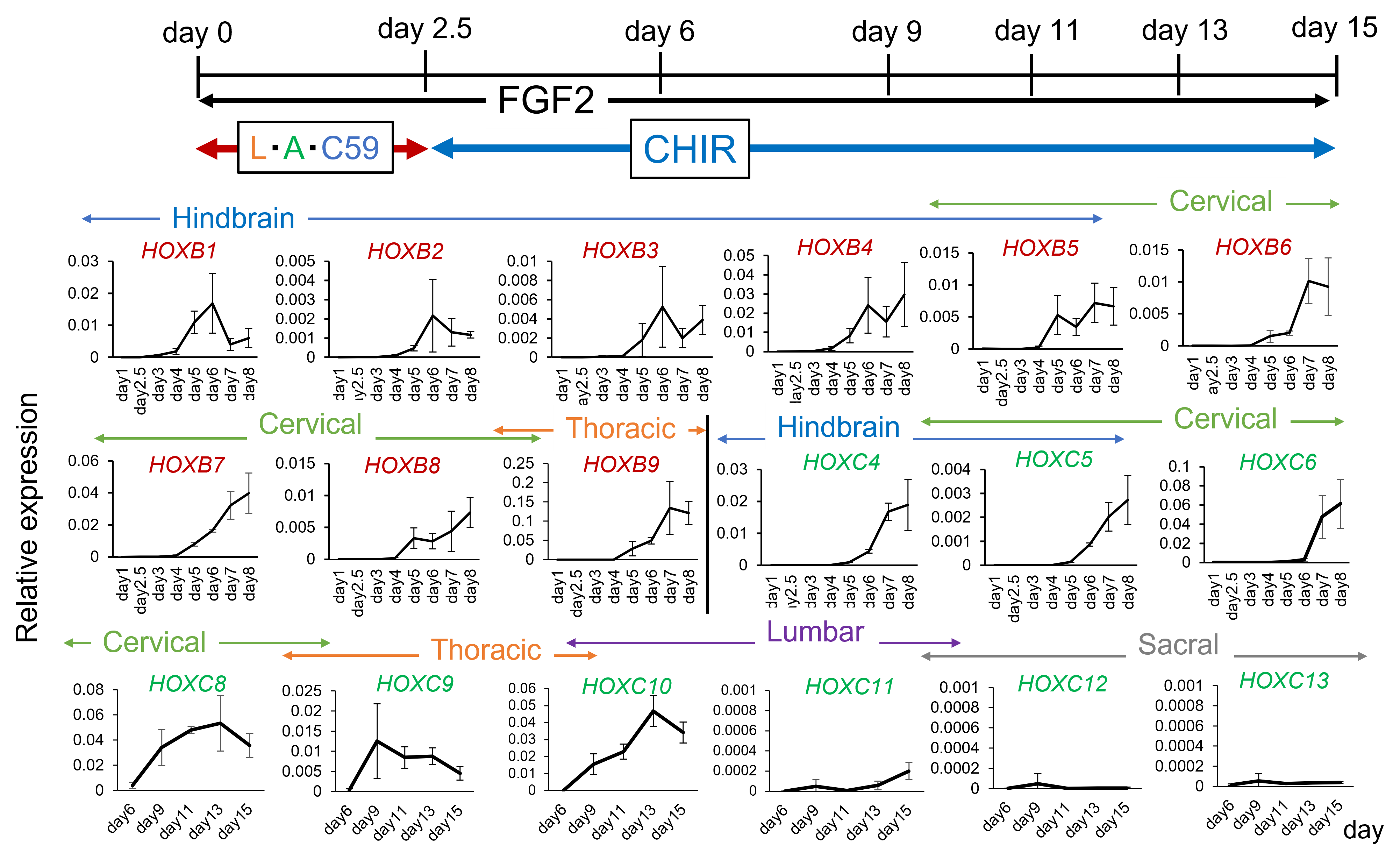
  • WNTシグナル活性化(CHIR)の添加タイミングの条件検討により、NMPsの高効率な誘導に成功(Fig.1)
  • 同一の培養系において健常iPS細胞・疾患ALS/CMS iPS細胞を用いて、胸髄領域におけるNMJの誘導に成功(Fig.3)
  • 誘導されたNMJは、クラーレまたはα-ブンガロトキシンの添加により筋収縮が低下

神経中胚葉前駆細胞の効率的な誘導法

Fig.1) Optimizing WNT activation timing efficiently induced NMPs : Exposing cells to CHIR from days 2-2.5 of differentiation efficiently induced cells that co-expressed the NMP markers SOX2 and TBXT. (n=3) LDN : BMP inhibitor, C59: WNT inhibitor.

胸髄運動領域、骨格筋前駆細胞の誘導確認

Fig. 2) The thoracic spinal motor region was induced in a concentration-dependent manner by SAG, SHH agonist : By increasing concentration of SAG, SHH agonist, expression of the motor region (OLIG2) was confirmed. Furthermore, under 150 nM SAG condition, expression of PAX3 in skeletal muscle progenitor cells was also detected (the neural marker SOX2 was negative). SB: TGF-b inhibitor, EC23: retinoic acid agonist, L: LDN, A: Activin.

NMJの構成因子の発現を確認

Fig. 3) Formation of the NMJ by maturation of motor neurons and skeletal muscles : From day 18, cells were attached and cultured in wells coated with laminin, poly-L-ornithine, and fibronectin. Muscle contraction was observed from around day 30. Expression of motor neurons (ISL1, TUJ1) and skeletal muscle (Titin, Myosin heavy chain; MHC) markers was confirmed at day 50. α-Bungarotoxin; Acetylcholine receptor marker.

分化誘導過程における脊髄部位の誘導変化

Fig. 4) FGF2 and CHIR induced the anterior-posterior spinal cord regions : Under this culture conditions (FGF2 20 ng/ml, CHIR 3 µM), induction of the spinal cord progressed over time and reached the lumbar region (HOXC10). (n=3)

応用可能性

● 頚髄・腰髄領域へのNMJ誘導法の拡張(Fig.4)
● NMJ関連疾患患者由来iPS細胞を用いて、疾患メカニズムの解明や新規治療薬開発への活用
● 老化・サルコペニア研究や神経・筋の部位特異的疾患への応用

特許

  • 関連特許出願中

← 研究シーズ&ニーズ集の一覧ページへ戻る


← 研究シーズ&ニーズ集のトップページへ戻る